lateral foot x ray anatomy
Radiographic Anatomy of the Skeleton: Cervical Spine -- Anteroposterior. 9 Pics about Radiographic Anatomy of the Skeleton: Cervical Spine -- Anteroposterior : Lisfranc fracture-dislocation | Image | Radiopaedia.org, Radiographic Anatomy of the Skeleton: Ankle -- Mortise View, Labelled and also Hip Xray | eORIF.
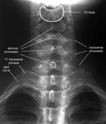
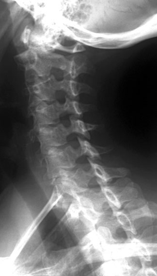
Radiographic Anatomy Of The Skeleton: Cervical Spine -- Anteroposterior
 uwmsk.org
spine ray ap cervical labelled neck anatomy xray normal anteroposterior radiology anterior radiographic spinal skeleton uwmsk injuries cspine posterior oblique
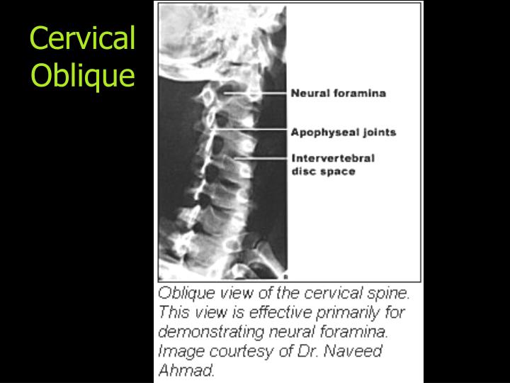
PPT - Normal X-Ray Anatomy PowerPoint Presentation - ID:867049
 www.slideserve.com
oblique cervical
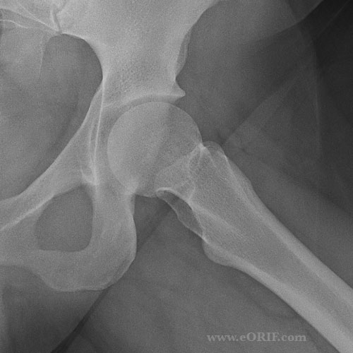
Hip Xray | EORIF
 eorif.com
hip xray lateral joint lat arthritis fracture neck eorif femoral head cysts narrowing ddh avn evaluate scfe osteophytes helpful etc
Congenital Vertical Talus | Image | Radiopaedia.org
 radiopaedia.org
talus vertical congenital foot rocker bottom radiopaedia lateral radiology ray case version calcaneus
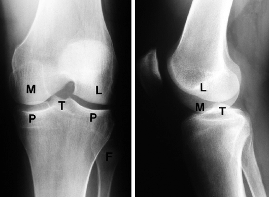
Applied Anatomy Of Knee Joint | Epomedicine
 epomedicine.com
knee anatomy joint lateral ap ray condyle xray femoral medial applied epomedicine radiographic
Lisfranc Fracture-dislocation | Image | Radiopaedia.org
 radiopaedia.org
lisfranc fracture dislocation radiopaedia radiology traumatic radiographs emrad homolateral
Radiographic Anatomy Of The Skeleton: Ankle -- Mortise View, Labelled
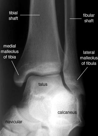 uwmsk.org
mortise ankle radiology anatomy labelled radiographic foot labeled lateral oblique medical talus ortopedia anatomical unlabelled version
Tarsal Coalition | Image | Radiopaedia.org
 radiopaedia.org
coalition tarsal talocalcaneal calcaneonavicular radiopaedia wikidoc version
Radiographic Anatomy Of The Skeleton: Cervical Spine -- Left Anterior
 uwmsk.org
spine cervical ray oblique labelled anatomy radiology left anterior neck radiographic skeleton diagram normal imaging vertebrae head uwmsk tech xray
Hip xray lateral joint lat arthritis fracture neck eorif femoral head cysts narrowing ddh avn evaluate scfe osteophytes helpful etc. Applied anatomy of knee joint. Tarsal coalition