leg vascular anatomy
Veins of the Lower Limb - Anatomy | Kenhub. 9 Images about Veins of the Lower Limb - Anatomy | Kenhub : Upper Extremity Venous Doppler – Sonographic Tendencies | Vascular, Upper Extremity and also Veins of the Lower Limb - Anatomy | Kenhub.
Veins Of The Lower Limb - Anatomy | Kenhub
vein kenhub lower veins femoral limb anatomy femoralis vena muscle artery nerve major
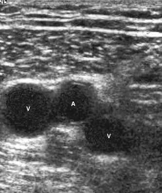
Upper Extremity Venous Doppler – Sonographic Tendencies | Vascular
 www.pinterest.com
ultrasound extremity venous doppler vein vascular sonographic thrombosis tendencies embolism artery sonographictendencies походження піна
Anatomy Of The Lower-limb Venous System And Assessment Of Venous
 radiologykey.com
vein artery lower femoral venous bifid duplicated anatomy transverse limb ultrasound assessment veins system insufficiency paired lying between radiology figure
Tennis Leg | Image | Radiopaedia.org
 radiopaedia.org
tennis leg ultrasound radiopaedia version
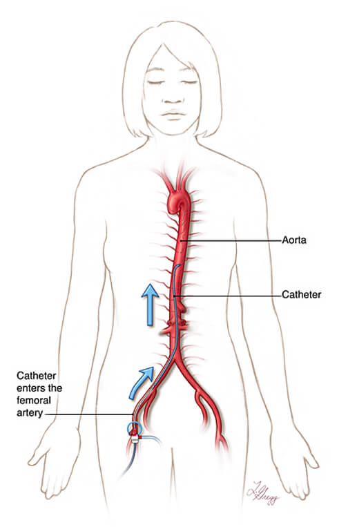
Spinal Angiography, Johns Hopkins Interventional Neuroradiology
 www.hopkinsmedicine.org
spinal angiography pathway spine femoral catheter artery aorta path hopkinsmedicine illustration neuroradiology interventional diagnostic figure johns hopkins arteries
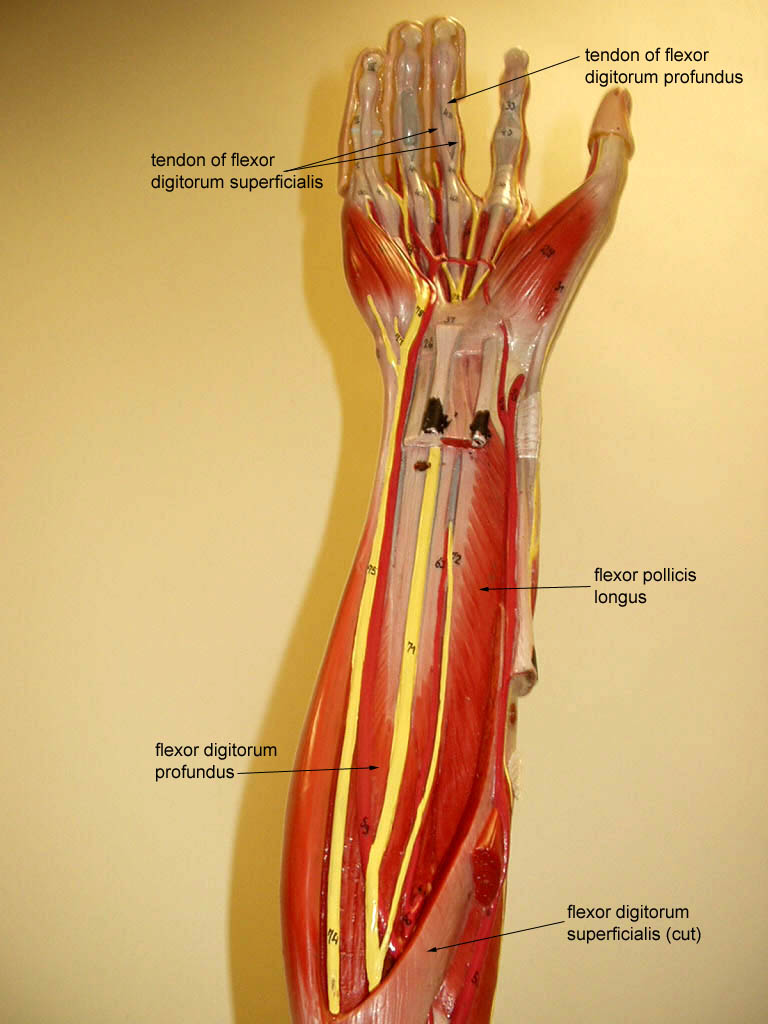
Upper Extremity
 classroom.sdmesa.edu
upper anatomy extremity physiology labeled arm muscles muscle lab label somso models human classroom biology deep nerve forearm anterior sdmesa
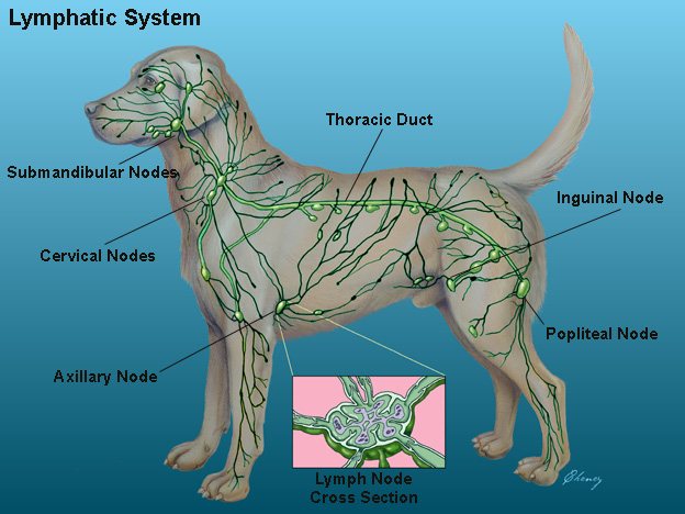
Glendale Animal Hospital - Veterinarian In Glendale, AZ USA :: Dog
 familyvet.com
system lymphatic anatomy dog canine chien cat lymph animal anatomie nodes animals vet health systems dogs du immune medicine chat
Image | Radiopaedia.org
 radiopaedia.org
cyst radiopaedia posttraumatic modality
Bipartite Hallux Sesamoid | Image | Radiopaedia.org
 radiopaedia.org
bipartite hallux sesamoid rays foot feet left medial metatarsal tibial head radiopaedia ray normal ankle sesamoids radiology anatomy case 1st
Bipartite hallux sesamoid rays foot feet left medial metatarsal tibial head radiopaedia ray normal ankle sesamoids radiology anatomy case 1st. Glendale animal hospital. Spinal angiography, johns hopkins interventional neuroradiology