long bone anatomy trabecula
Schematic diagram of a portion of a long bone showing the articular. 9 Pictures about Schematic diagram of a portion of a long bone showing the articular : This illustration shows the spongy bone within the proximal epiphysis, Adult long bone. Sagittal section through long bone showing the and also Schematic diagram of a portion of a long bone showing the articular.
Schematic Diagram Of A Portion Of A Long Bone Showing The Articular
 www.researchgate.net
articular trabecular cartilage cortical medullary cavity periosteum schematic
This Illustration Shows The Spongy Bone Within The Proximal Epiphysis
 oerpub.github.io
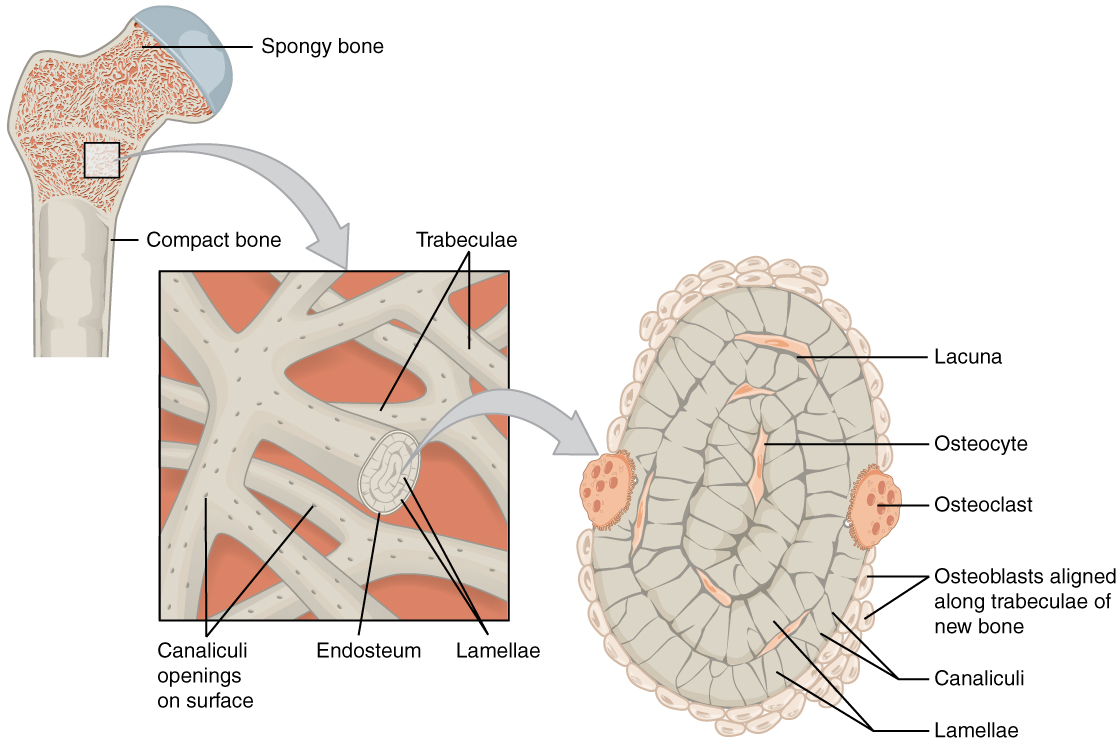
bone spongy trabeculae cancellous compact endosteum cells within canaliculi osteocytes called lamellae lacunae marrow
Adult Long Bone. Sagittal Section Through Long Bone Showing The
 www.researchgate.net
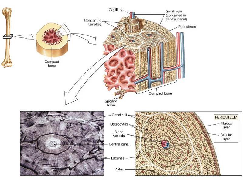
sagittal cancellous cortex cortical spongy trabeculae spicules skeletal marrow endosteum abdelmagid samir
Bone And Osteocytes - Essentials Of Anatomy And Physiology
 essentialsofanatomyandphysiology.weebly.com
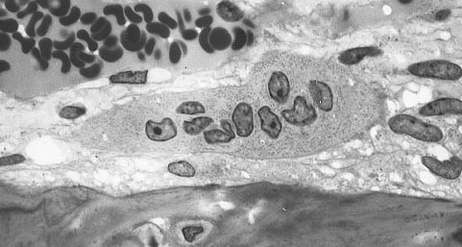
osteocytes osteoclasts
Osteon Diagram Lab | Wiring Diagram
 14.yoga-neuwied.de
osteon spongy trabeculae osteons parallel histology
Long Bone Cross Section / The Parts Of A Healthy Long Bone With A Cross
 drawingoutthedepths.blogspot.com
cortical europepmc blood interstitial capillary diaphysis arterial dense
Bone And Osteocytes - Essentials Of Anatomy And Physiology
 essentialsofanatomyandphysiology.weebly.com
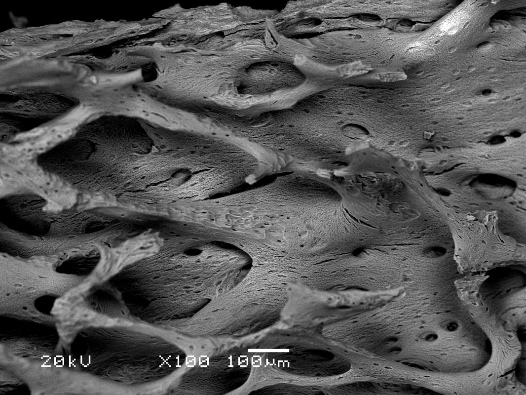
trabecular microscopic knochen trabeculae medicalnewstoday x100 osteocytes gereken bilmeniz kemikler musst devez formed magnification cancellous demedbook wistar bertazzo tif rat
Anatomy Exam 1 Flashcards | Easy Notecards
 www.easynotecards.com
bone compact canal central anatomy cortical lacuna exam lamella canaliculus easynotecards
33 Label The Structures Of The Bone Using The Hints Provided. - Labels
 opilizeb.blogspot.com
Sagittal cancellous cortex cortical spongy trabeculae spicules skeletal marrow endosteum abdelmagid samir. Bone compact canal central anatomy cortical lacuna exam lamella canaliculus easynotecards. Osteocytes osteoclasts